Course Description: Understanding the scope and significance of neuroimaging.
Definition and Scope:
What is Brain Mapping? Brain mapping is a comprehensive process of identifying the physical structure and functional properties of various brain regions, including their connections.
What is Neuroimaging? Neuroimaging encompasses a suite of non-invasive imaging techniques that allow visualization and measurement of brain structure, activity, and metabolism.
Structural vs. Functional Imaging:
| Structural Imaging | Functional Imaging |
| MRI and CT scans for detailed anatomical visualization. | fMRI, PET, and SPECT for observing dynamic processes like blood flow and metabolic activity |
The Scope of Neuroimaging:
Applications in healthcare, cognitive neuroscience, and advanced research fields such as AI and brain-computer interfaces.
Applications:
| In Clinical Diagnosis: | In Research: | In Research: |
| Detecting and monitoring diseases like Alzheimer’s, epilepsy, and multiple sclerosis. | Unraveling the neural mechanisms behind memory, emotion, and decision-making. | Guiding neurosurgical procedures with precision. |
| Identifying tumors, vascular anomalies, and traumatic brain injuries. | Understanding developmental changes and age-related neurodegeneration. | Enhancing outcomes through personalized approaches. |
Real-World Example:
A notable example of neuroimaging leading to groundbreaking discoveries is the development of a rapid diagnostic test for medulloblastoma, the most common brain tumor in children. Researchers from the University of Birmingham and Newcastle University utilized advanced MRI scans combined with artificial intelligence to identify specific chemical signatures of the tumor. This innovative approach reduced the diagnostic time from several weeks to just ten minutes, enabling quicker and more targeted treatments, thereby significantly improving patient outcomes.
Read the full article here.
Techniques Overview:
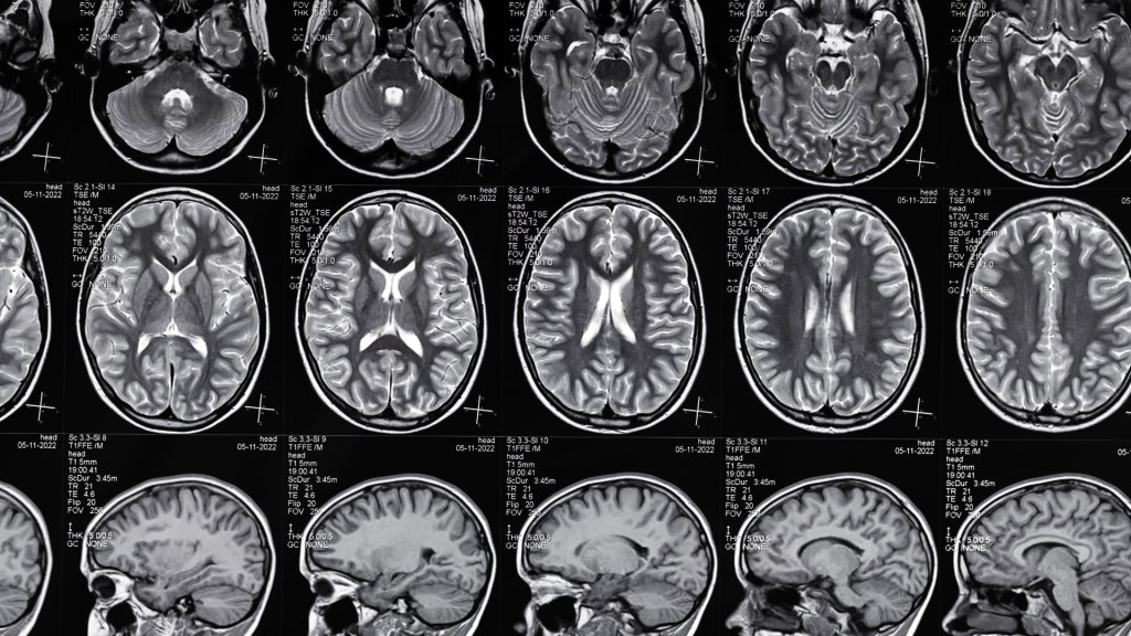
MRI (Magnetic Resonance Imaging):
- High-resolution images of brain structure.
- Applications: Tumor detection, stroke assessment, and congenital abnormalities.
CT (Computed Tomography):
- Quick imaging for acute conditions like hemorrhages and fractures.
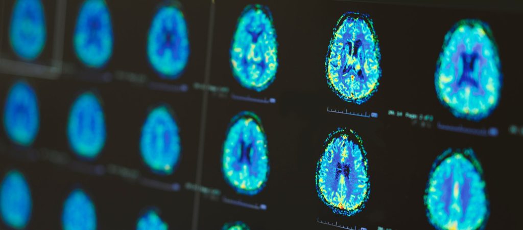
PET (Positron Emission Tomography):
- Insight into metabolic activity and receptor functionality.
- Applications in oncology, cardiology, and neurology.
fMRI (Functional MRI):
- Measures BOLD (Blood-Oxygen-Level-Dependent) signals.
- Applications in understanding functional connectivity and brain activation patterns.
| Technique | Resolution | Cost | Primary Use Cases |
| MRI | High | Moderate to High | Structural imaging for tumors, strokes |
| CT | Moderate | Low | Quick imaging for acute conditions |
| PET | Moderate | High | Quick imaging for acute conditions |
| fMRI | High | High | Functional connectivity and brain activation |
Impact on Neuroscience:
Revolution in Diagnoses:
- Shift from symptomatic diagnosis to biomarker-based identification of diseases.
Advancements in Treatment:
- Development of targeted therapies for neurological and psychiatric conditions.
- Role in drug development through understanding disease mechanisms.
Broader Implications:
- Insights into brain plasticity and recovery after injury.
- Contribution to the fields of neuroethics and human enhancement.
Case Studies:
1. Detecting Residual Cognitive Function in Patients with Severe Brain Injury
Traditional behavioral assessments often misdiagnose patients in vegetative or minimally conscious states, with error rates up to 43%. Advanced neuroimaging techniques, such as functional MRI (fMRI) and electroencephalography (EEG), have been employed to detect residual cognitive function in these patients. Studies have shown that a subset of patients diagnosed as vegetative retain conscious awareness undetectable through bedside examinations. In some cases, neuroimaging has facilitated communication with these patients, enabling them to participate in decisions about their care. This approach has improved diagnostic accuracy and informed treatment strategies, significantly impacting patient outcomes.
Read the full study here.
2. Understanding Aphantasia: The Absence of Mental Imagery

Aphantasia is a condition where individuals cannot visualize images in their minds. Neuroimaging studies have investigated the neural basis of this phenomenon, revealing that people with aphantasia exhibit different patterns of brain connectivity, particularly between the prefrontal cortex and visual processing areas. These findings suggest that the lack of mental imagery in aphantasia results from reduced communication between brain regions responsible for generating and integrating visual information. This research has deepened scientific understanding of the diversity in human cognition and perception, highlighting the neural mechanisms underlying mental imagery.
Read the full study here.