Innovations in Ultra-High-Resolution Imaging
Innovations in Ultra-High-Resolution Imaging: Developments in 7T MRI and Beyond
Objective:
To explore the innovations in ultra-high-resolution neuroimaging, focusing on developments in 7T MRI and its potential advancements.
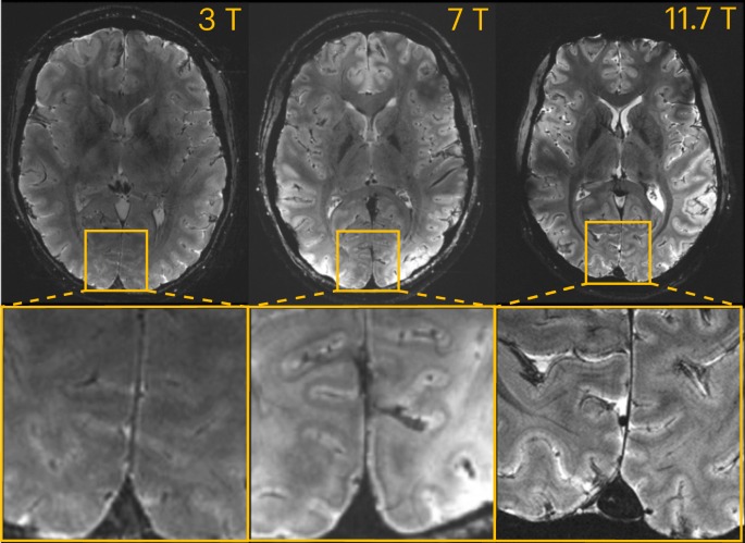
Ultra-high-resolution imaging refers to the ability to capture brain images at a much finer level of detail than conventional MRI techniques. Magnetic Resonance Imaging (MRI) is already an essential tool in neuroimaging, but developments in ultra-high-field MRIs, particularly the 7T MRI (7 Tesla), are pushing the boundaries of what can be visualized in the brain. These advancements allow for more precise images of the brain’s structure, enabling researchers and clinicians to identify subtle abnormalities that may not be visible with standard MRI scans.
Key Concepts in Ultra-High-Resolution Imaging:
- 7T MRI:
- A 7 Tesla MRI offers a significantly stronger magnetic field than traditional MRI scanners (1.5T or 3T). This increased field strength enhances the signal-to-noise ratio, enabling clearer and more detailed images, particularly in areas like the cortical gray matter and deep brain structures.
2. Sub-millimeter Resolution:
- Ultra-high-field MRIs can achieve resolution at the sub-millimeter level, which allows for the visualization of microstructures, such as individual layers of the cortex and finer white matter tracts.
3. Improved Functional Imaging:
- In addition to structural imaging, 7T MRI also improves functional MRI (fMRI) sensitivity, enabling better detection of brain activity and more precise mapping of brain regions involved in specific tasks.
Applications of Ultra-High-Resolution Imaging:
Detailed Brain Mapping:
- 7T MRI enables the creation of detailed, high-resolution brain maps, which can be used to better understand the organization of different brain areas, both structurally and functionally.
Neurodegenerative Diseases:
- High-resolution imaging allows for early detection of subtle changes in brain regions affected by neurodegenerative diseases like Alzheimer’s and Parkinson’s disease, providing an opportunity for early intervention.
Microstructural Abnormalities:
- The ability to visualize fine structures means that 7T MRI can detect early-stage changes in diseases that affect small areas of the brain, such as multiple sclerosis and schizophrenia.

Real-World Example:
Brain Tumor Visualization:
- 7T MRI has been used in research and clinical settings to visualize brain tumors with much higher clarity than traditional MRI. This allows for a more accurate delineation of tumor boundaries and better planning for surgical interventions.
Case Study:
Epilepsy:
- In patients with drug-resistant epilepsy, 7T MRI can identify subtle structural abnormalities that traditional MRI may miss. This detailed imaging is particularly valuable when localizing seizure foci for surgical planning.